
My lab is a member of the ES Cell Research Group at Washington University. A major goal of our work is to shed light on the factors that control neuronal differentiation. An embryo starts out as a single totipotent stem cell, but quickly gives rise to thousands of distinct cell types including many different kinds of neurons and glial cells. Recent work from a number of laboratories, including Dave Gottlieb's group at Washington University, has shown that multipotential stem cells can be induced to differentiate into neurons in vitro following exposure to retinoic acid. We are studying the physiological properties of neurons that arise from Embryonic Stem (ES) cells and from the P19 embryonic carcinoma cell line.
Our initial goal has been to determine how far these cells can progress along the pathway of neuronal differentiation. To date, we have shown that both P19- and ES-derived neurons express voltage-gated sodium, potassium, and calcium channels, as well as channels gated by the neurotransmitters glutamate, GABA, and glycine. Within 2 to 3 weeks after induction by retinoic acid, some of the cells have formed excitatory synapses, whereas others have formed inhibitory connections. Using antibodies raised against growth associated protein-43 (GAP43) and microtubial associated protein-2 (MAP-2) we have demonstrated the segregation of these proteins into separate axonal and somatodendritic compartments, respectively. We also showed that a number of synaptic vesicle antigens, including synapsin, synaptophysin and SV2, are localized to discrete sites of synaptic contact. In both their electrophysiological properties and their ability to establish a polarized phenotype, therefore, these cells strongly resemble neurons from the CNS.


We are currently testing whether exposure to additional growth and differentiation factors can influence the mature phenotye attained by these cells. In addition we are using molecular biology to alter the expression of proteins that are thought to be involved in synaptic transmission.
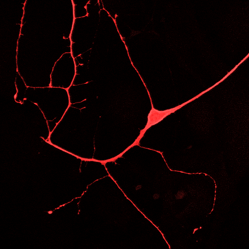
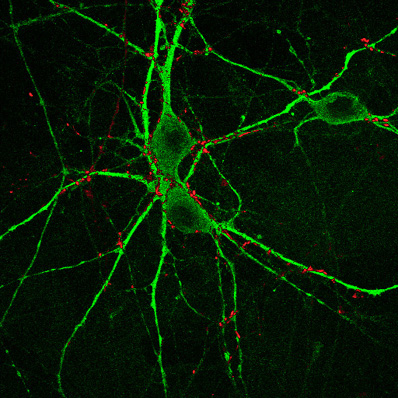
The second image on this page shows an ES cell in culture that was labeled with DiI.
The third image on this page shows a P19 cell culture that was double labeled using antibodies to MAP-2 (green) and to the vesicle protein synapsin (red).
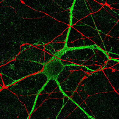
The image on our home page shows a P19 cell culture that was double labeled using antibodies to the proteins GAP43 (red) and MAP2 (green).
 Return to the Huettner Lab Home Page